Anatomy Of Chest - Anatomy Chapter 10 11 Frontal Chest Muscles Diagram Quizlet / Normal anatomic structures are labeled on posteroanterior (pa) and lateral chest radiographs (figs.
Anatomy Of Chest - Anatomy Chapter 10 11 Frontal Chest Muscles Diagram Quizlet / Normal anatomic structures are labeled on posteroanterior (pa) and lateral chest radiographs (figs.. The chest anatomy includes the pectoralis major, pectoralis minor and the serratus anterior. The embryologic and anatomic basis of modern surgery. Anatomy of the chest and the lungs: Normal anatomic structures are labeled on posteroanterior (pa) and lateral chest radiographs (figs. Is its effect so thoroughly nebulous that it's hard to justify?
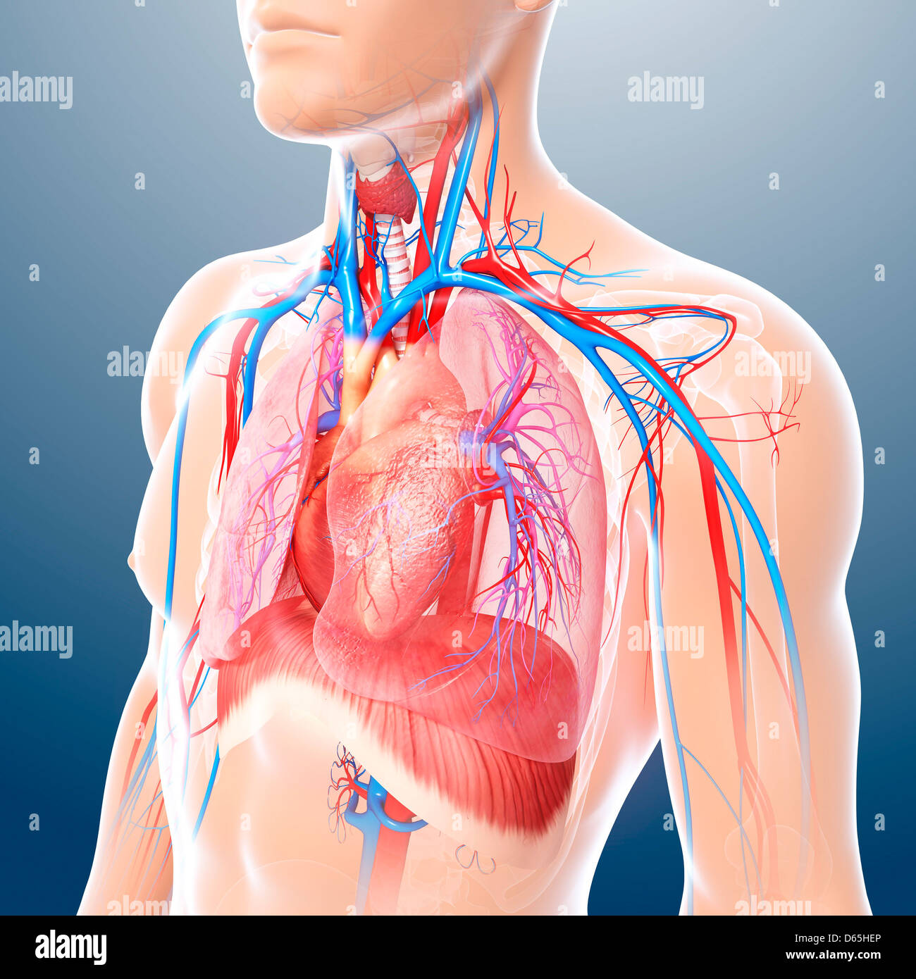
The chest anatomy includes the pectoralis major, pectoralis minor and the serratus anterior. Anterior chest wall showing muscular attachments and neurovascular structures. The chest is the area of origin for many of the body's systems as it houses organs such as the heart, esophagus, trachea, lungs, and the circulatory system does most of its work inside the chest. Book of chest anatomy is a passive item. Here's how science can help you grow!► get the full built by science program.
The first is the pectoralis major which is the largest one and located in the center of the chest.
The first is the pectoralis major which is the largest one and located in the center of the chest. Is its effect so thoroughly nebulous that it's hard to justify? 12 photos of the anatomy of the chest. Fill out your shirt with a bigger, stronger, more powerful chest. Find out more about the individual muscles. Anatomy is to physiology as geography is to history: Choose from 500 different sets of flashcards about chest anatomy on quizlet. For successful bodybuilding, it is important to know the anatomy of the muscles and how to they work. Book of chest anatomy is a passive item. Set 5 imaging tutorial #2; Anterior chest wall showing muscular attachments and neurovascular structures. This mri chest (thorax) axial cross sectional anatomy tool is absolutely free to use. It describes the theatre of events.
Radiology basics of chest ct anatomy with annotated coronal images and scrollable axial images to help medical students and junior doctors learning anatomy. Find out more about the individual muscles. Here's how science can help you grow!► get the full built by science program. Learn about chest anatomy with free interactive flashcards. Anatomy is to physiology as geography is to history:

The chest wall is supplied by the posterior intercostal arteries arising from the aorta, the internal thoracic and the.
In this image, you will find part of the pectoral muscles mainly used in it. The embryologic and anatomic basis of modern surgery. Is its one synergy actually worthwhile? The chest anatomy includes the pectoralis major, pectoralis minor and the serratus anterior. Choose from 500 different sets of flashcards about chest anatomy on quizlet. Fill out your shirt with a bigger, stronger, more powerful chest. Radiology basics of chest ct anatomy with annotated coronal images and scrollable axial images to help medical students and junior doctors learning anatomy. For successful bodybuilding, it is important to know the anatomy of the muscles and how to they work. This mri chest (thorax) axial cross sectional anatomy tool is absolutely free to use. This page provides an overview of the chest muscle group. Normal anatomic structures are labeled on posteroanterior (pa) and lateral chest radiographs (figs. Book of chest anatomy is a passive item. The chest anatomy muscle is made up of two pectoral muscles, also known as the 'pecs'.
This mri chest (thorax) axial cross sectional anatomy tool is absolutely free to use. The embryologic and anatomic basis of modern surgery. Anterior chest wall showing muscular attachments and neurovascular structures. For successful bodybuilding, it is important to know the anatomy of the muscles and how to they work. 12 photos of the anatomy of the chest.
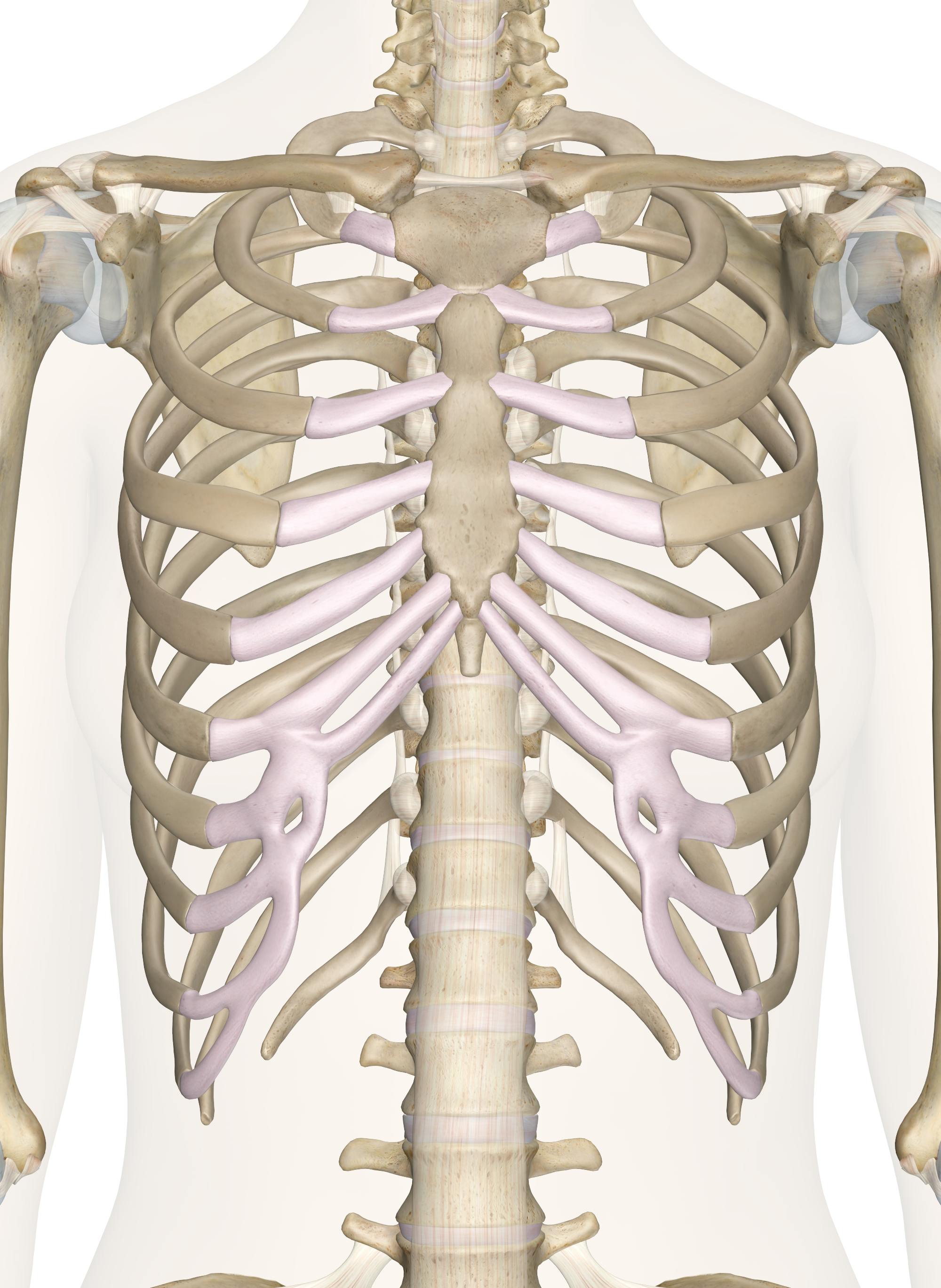
Basic rib anatomy consists of a head, neck, tubercle, angle, shaft, and costal groove.
Sectional anatomy of the the chest.— 3 window levels there are certain structures within the chest which require specific density and contrast settings known as. Normal anatomic structures are labeled on posteroanterior (pa) and lateral chest radiographs (figs. The embryologic and anatomic basis of modern surgery. The first is the pectoralis major which is the largest one and located in the center of the chest. Improves the contents of broken chests. The chest anatomy includes the pectoralis major, pectoralis minor and the serratus anterior. Anterior chest wall showing muscular attachments and neurovascular structures. Set 5 imaging tutorial #2; This page provides an overview of the chest muscle group. The chest wall is supplied by the posterior intercostal arteries arising from the aorta, the internal thoracic and the. Anatomy is to physiology as geography is to history: Sixteen + sets of chest exercises with very little or no results aside from pain and tenderness within the front of. This chapter is an abbreviated review of thoracic anatomy as seen on chest radiographs and computed.


Komentar
Posting Komentar